
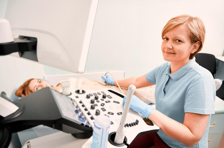
Sonography is a type of diagnostic imaging that allows medical professionals to view different body areas to diagnose and monitor a range of illnesses in adults, children, and women. Sonography, often known as ultrasound, uses sound waves to reach the area of the body that the physician wants to examine.
The sonographer’s duties include:
- Taking the images.
- Showing them to the doctor or other medical professional.
- Interacting with the patient throughout the imaging process.
- Supporting the identification of abnormalities.
A career as a sonographer, an ultrasound technician, or a diagnostic medical sonographer is exciting and rewarding.
Here, we are going to discuss different types of sonographers.
1.Obstetric Sonographer for Obstetrical Ultrasound of Fetuses
The specialty of obstetric sonographers is imaging fetuses throughout pregnancy. Obstetric sonographers can monitor a fetus’s growth and development through sonograms, which can assist doctors in creating health procedures that women can follow throughout their pregnancies.
Moreover, obstetric sonographers can obtain pictures of the uterus to figure out a woman’s pregnancy status and estimate the baby’s due date. Most obstetric sonographers’ careers are in medical centers and hospitals with maternity wards.
It is excellent, great news. If your pregnancy test results are positive, visit a well-reputed clinic such as obstetrical ultrasound marietta ga in Marietta, GA, clinic for a features ultrasound.
2.Vascular Sonographers for Ultrasound of Blood Vessels
A vascular technologist, a vascular sonographer, takes images of your blood vessels, such as your arteries and veins. They also gather information that contributes to medical professionals’ diagnosis of blood flow-related disorders. Vascular technologists regularly examine your blood flow to identify blockages in arteries or blood clots.
3.Diagnostic Cardiovascular Sonographer for Heart Problems
Cardio sonographers, also known as echocardiographers, assist physicians in diagnosing heart problems by working with cardiac specialists. The apparatus uses two and three-dimensional images of the heart and its chambers.
To check for blockages or cardiac muscle damage, doctors examine the blood flow and anatomy of the heart. Doctors can recommend medication, surgery, or lifestyle modifications based on the images they see to stop more heart damage.
4.Musculoskeletal Sonographer for Ultrasound of Muscles and Skeletal System
A musculoskeletal sonographer uses imaging to diagnose muscle and skeletal system component problems. This covers every tendon, ligament, joint, and nerve in the patient’s body. Sonographers who specialize in the musculoskeletal system often use the images they take to identify conditions that impact a patient’s joints, muscles, and bones and can, therefore, limit their mobility.
Furthermore, musculoskeletal sonographers frequently work in emergency rooms, hospitals, and other medical facilities that provide care for patients with injuries caused by trauma. When doing their work, musculoskeletal sonographers watch out for the following conditions: strains, arthritis, hernias, cysts, tense muscles or items, bone fractures, sprains, inflammation, and strains.
5.Breast Sonographer
A breast sonographer is an expert in capturing diagnostic images of the breast and surrounding tissue following an abnormal mammography or examination. They often perform imaging of the breasts, tissue, and lymph nodes as part of their work to search for signs of a developing medical illness.
Among the abnormalities breast sonographers commonly search for inpatients are lumps, cysts, and tumors. Breast sonographers can help physicians diagnose cancer as well, and the images they acquire may reveal areas that indicate the possibility of malignant growth.
6.Ophthalmic Sonographers for Ultrasound of Eyes
Sonographers specializing in ophthalmology use ultrasound to examine the eyes and surrounding tissues. They generate images of the inside structures of the eye using sound waves. Sonographers look at organs such as the optic nerve, vitreous fluid, cornea, and lens.
These specialists search for anomalies, growths, foreign objects, retinal detachments, and other diseases. Ophthalmic sonographers may choose different specializations. They may focus on pediatric ophthalmic ultrasonography, glaucoma evaluation, or oncology.


More Stories
The Ultimate Work Desk Kit: From Eye Drops to Vicks Roll On
Essential Insights into Atlanta Insurance: Your Ultimate Guide
Transform Lives by Giving Plasma: A Comprehensive Guide